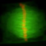
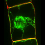
 Division of tobacco suspension-cultured cell.
 Orange and green colors represent chromosomes and microtubules, respectively. Movie shows 600 times faster than real time. (Provided by Drs. Tomomi Hayashi and Sei-ichiro Hasezawa in The University of Tokyo)
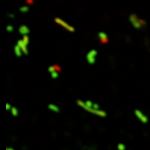 Fusion of mitochondria in onion cells.
 Distinct mitochondria are visualized with red and green colors, respectively. Fusion between two mitochondria results in yellow color. Movie shows 30 times faster than real time.
(Provided by Dr. Shin-ichi Arimura in The University of Tokyo)
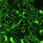 Movement of ER and ER bodies.
 When GFP was used to visualize ER, rod-shape structures were observed in addition to the typical network structure ER. The former and the latter represent ER bodies and ER, respectively. In this movie, both structures move dynamically in the cell. Movie shows 19 times faster than real time.
(Provided by Dr. Atsushi J. Nagano in Kyoto University)
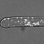 Movement of peroxisomes in root hair cell.
Peroxisomes in Arabidopsis root hair cells move frequently. Movie shows 10 times faster than real time.
(Provided by Dr. Shoji Mano in National Institute for Basic Biology)
 Division of tobacco suspension-cultured cell.
 Green and red colors represent endoplasmic reticulum and endosome, respectively. Cell plate formation is observed during cell division, because substrates, which are transported via the endosome, are involved in cell plate formation. Movie shows 600 times faster than real time.
(Provided by Dr. Tomomi Hayashi in The University of Tokyo)
 Division of chloroplasts in the moss Physcomitrella patens.
 Chloroplasts in this movie are not visualized with fluorescent proteins such as GFP. Observation with light microscopy clearly reveals chloroplast division in the moss Physcomitrella patens. Movie shows 1,800 times faster than real time.
(Provided by Dr. Hiroyoshi Takano in Kumamoto University)
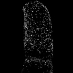 Distribution of peroxisomes in tobacco suspension-cultured cell.
 Peroxisomes are observed as spherical structures whose diameter is about 1 µm. Since the central vacuole is present in the center of the cell, peroxisomes are located in the narrow region between the central vacuole and plasma membrane, showing the morphology of the cell.
(Provided by Dr. Shoji Mano in National Institute for Basic Biology)
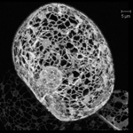 3D structure of ER network in tobacco suspension-cultured cell.
 The endoplasmic reticulum (ER) is observed as a network-like structure. Since the central vacuole is present in the center of the cell, this network-like structure is located in the narrow region between the central vacuole and plasma membrane, showing the morphology of the cell. The spherical structure in the cell is the nucleus, because the ER is connected with the nuclear membrane.
(Provided by Dr. Haruko Ueda in Kyoto University)
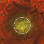 Avoidance movement of chloroplasts.
 Light is indispensable for plant growth. Chloroplasts are involved in photosynthesis using light. However, excess amount of light causes damage to plants. Therefore, chloroplasts escape from the cell surface under the strong light, which is represented by the circle in this movie.
(Provided by Dr. Aeko Kadota in Tokyo Metropolitan University)
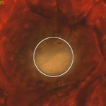 Accumulation movement of chloroplasts.
 In contrast to avoidance movement from strong light, chloroplasts accumulate at the cell surface to optimize photosynthesis under weak light, which is represent by the circle in this movie.
(Provided by Dr. Aeko Kadota in Tokyo Metropolitan University)