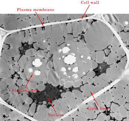
 The conditions inside plant cells are quite different among different tissues and developmental stages observed even in the same plant, as well as different plant species. The left picture shows an electron micrograph of cotyledonary cells of Arabidopsis seeds. The majority of the area in each cell is occupied with protein bodies and lipid bodies, both of which accumulate proteins and lipids, respectively. Immediately after germination, seedlings do not have the ability to undertake photosynthesis so they cannot produce energy by themselves. Therefore, they metabolize storage proteins and lipids in seeds to produce energy for post-germinative growth. At this time, the organelles have specific roles such as degradation of storage substrates.
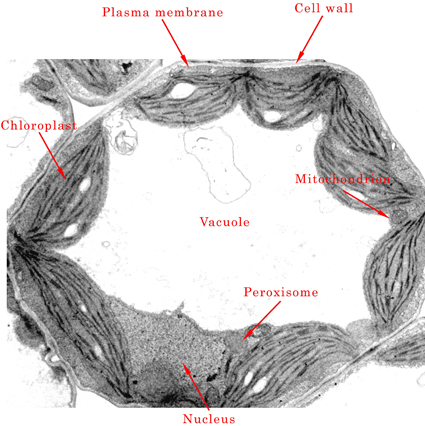 The right panel also shows Arabidopsis cotyledonary cells, but the content of cells is quite different from that of the previous picture. This picture was derived from green cotyledons whose greening has finished after germination. By this time, chloroplasts have been developed, and plants acquire the ability of photosynthesis. As a result, plants can produce energy from inorganic substrates. Therefore, various organelles in the cells change their functions to be suitable for photosynthesis. The central area in this picture represents the developed vacuole that contains various substrates such as proteins, inorganic compounds and dyes.